Sole Of Foot Diagram Ankle Tendons Ligaments Joint
Foot sole area measurement. the surface areas of 9 different individual Collection 100+ wallpaper anatomy of the bottom of the foot full hd, 2k Pin on anatomy reference
Foot Pain Diagram - Why Does My Foot Hurt? (2022)
Diagram of sole of foot Plantar fasciitis stretches: best stretches for fast relief Muscles of the foot laminated anatomy chart
Major structures of the sole of the foot, inferior view (right side
Ankle tendons ligaments jointHuman anatomy for the artist: the dorsal foot: how do i love thee? let Plantar foot anatomy nervesFoot diagram.
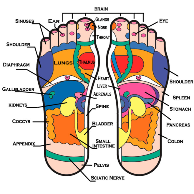
Pin on health picture referencesPlantar aspect of foot Shows the important areas / regions of the human foot bottomFeet reflexology body parts chart areas foot sole different massage simple haven part organs bottom other health organ map linked.

Diagram of your foot
Unveiling the perfect fit: debunking the myth of tight volleyball shoesSole of foot diagram Pressure massage reflexology acupuncture acupressure therapy healing shiatsu pain meridian musely oasis trivia involvesPlantar foot anatomy diagram.
Arteries quizlet suejeskekjFoot tendon ankle anatomy tendons diagram dorsal muscle lateral human hand muscles diagrams chart prp bone structure artist extensor me Foot sole feet innervation cutaneous wikipedia nerve diagram wiki svg pain nervesDiagram: muscles on sole of foot (2nd layer) diagram.

Hurt wiring
Susan's blog: feet haven reflexologyAnkle tendon calf ligament Medial muscles and bones of the foot sole labeled human anatomy diagramFoot pain diagram.
Sole (foot)Pressure point layout What is metatarsalgia?Foot anatomy bottom physiology using plantar structure netter bones medial surface layer superficial imagery relax notes weight arch inside aspect.

Lesser toe deformities — orthopaedicprinciples.com
Foot anatomy muscles and tendonsFracture metatarsalgia joionline joi Foot diagramSole of foot diagram.
Foot diagram with labelsFoot arteries diagram : arteries foot dorsal plantar view ankle stock Foot tendon anatomy diagramSoles individual toes male cutaneous digits.

A diagram of parts of the foot drawing by pat byrnes
Muscles of the foot photograph by asklepios medical atlas fine artTendons tendon deformities lesser orthopaedicprinciples 1135 Sole inferior structures muscles superficial layer diogoNotes on anatomy and physiology: using imagery to relax the weight.
.


